dCas9-mediated PCR-free Detection of Oncogenic Mutation by Non-equilibrium Nanoelectrokinetic Selective Preconcentration
Cutting-edge nanoelectrokinetic technology in this work provides a breakthrough for the present clinical demands of molecular diagnosis to detect a trace amount of oncogenic mutation of DNA in a short time without an erroneous PCR procedure. In this work, we combined the sequence-specific labeling scheme of CRISPR/dCas9 and ion concentration polarization (ICP) mechanism to separately preconcentrate target DNA molecules for rapid detection. Using the mobility shift caused by dCas9's specific binding to the mutant, the mutated DNA and normal DNA were distinguished in the microchip. Based on this technique, we successfully demonstrated the dCas9-mediated 1-min detection of single base substitution (SBS) in EGFR DNA, a carcinogenesis indicator. Moreover, the presence/absence of target DNA was identified at a glance like a commercial pregnancy test kit (two lines for positive and one line for negative) by the distinct preconcentration mechanisms of ICP, even at the 0.1% concentration of the target mutant.
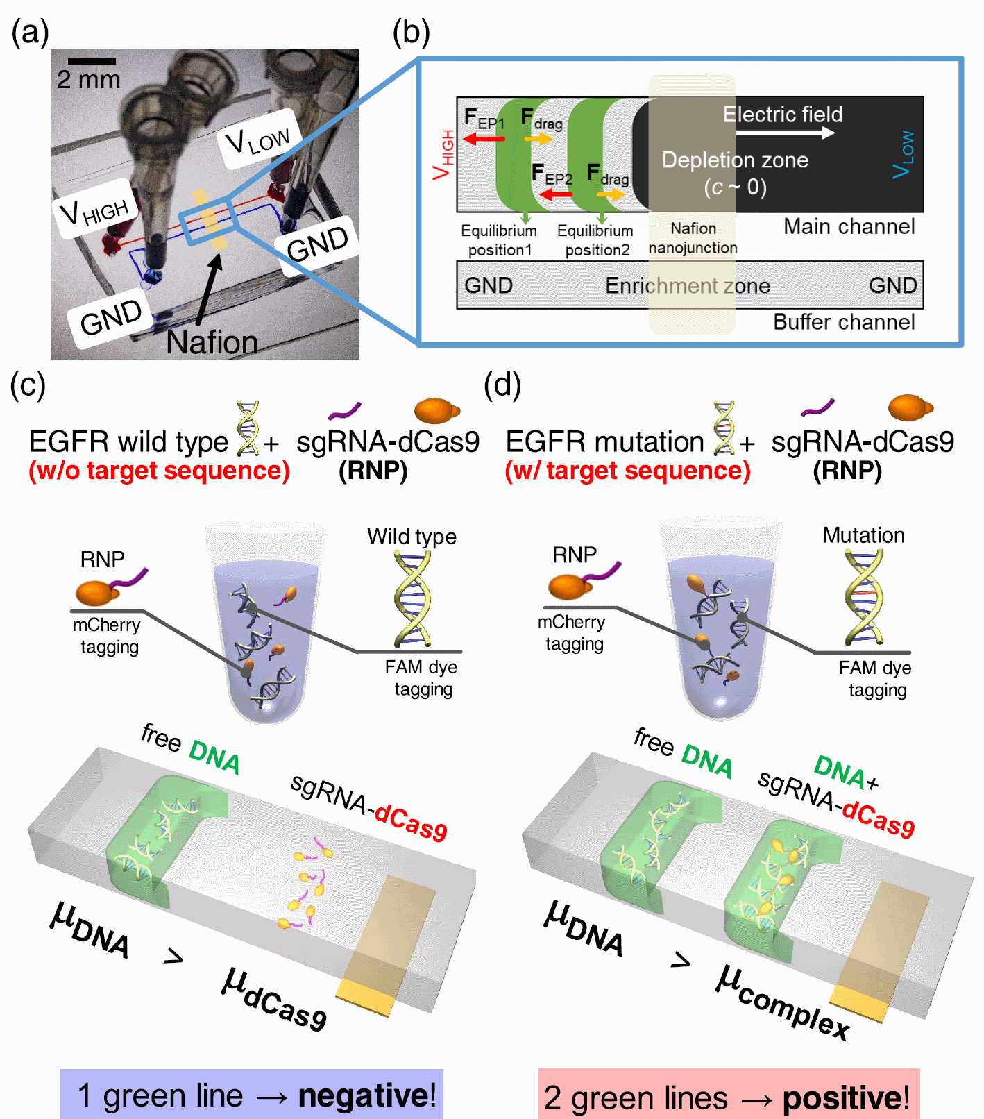
Figure 1. (a) Picture of a real micro/nanofluidic device. (b) Schematic diagram of preconcentration plugs with different balance positions. (c) Schematic of the negative case: RNP-targeting EGFRM was not bound to EGFRWT so that this case showed one green plug from EGFRWT. (d) Schematic of the positive case: RNP-targeting EGFRM was successfully bound to EGFRM so that there are two green plugs from the RNP + DNA complex and unbound DNA.
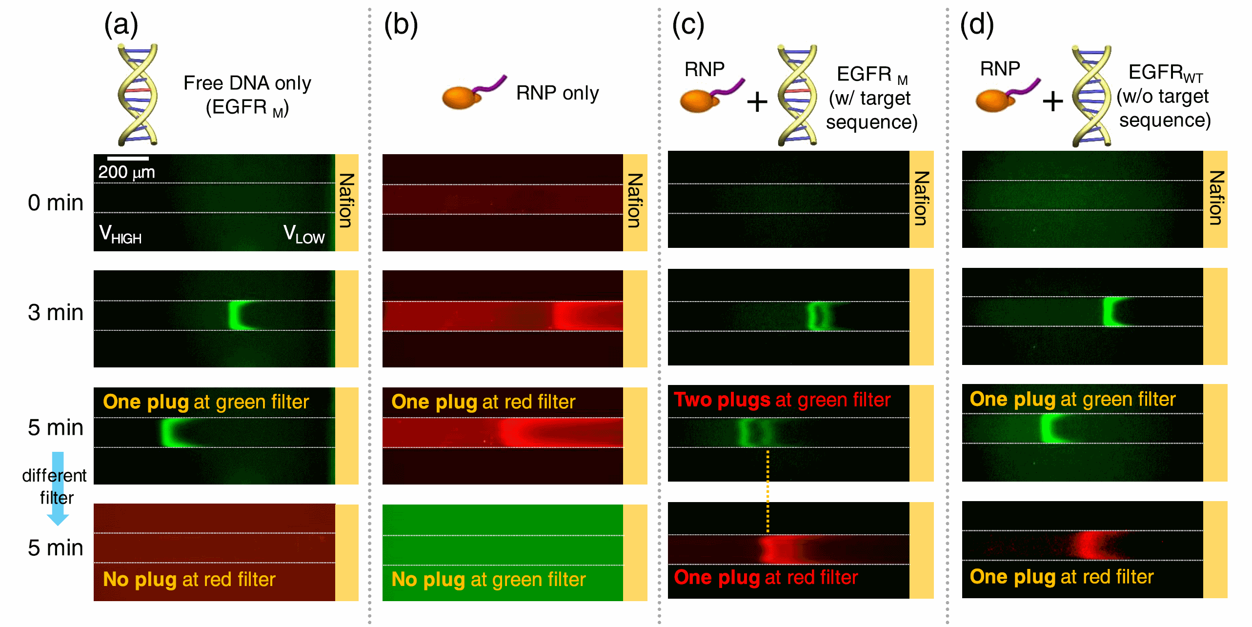
Figure 2. Fluorescence results of (a) free DNA, (b) RNP, (c) RNP with EGFRM, and (d) RNP with EGFRWT using a selective concentration of nanoelectrokinetics selective preconcentration. It was confirmed that RNP could be combined only with EGFRM to detect SBS in the micro/nanofluidic platform.
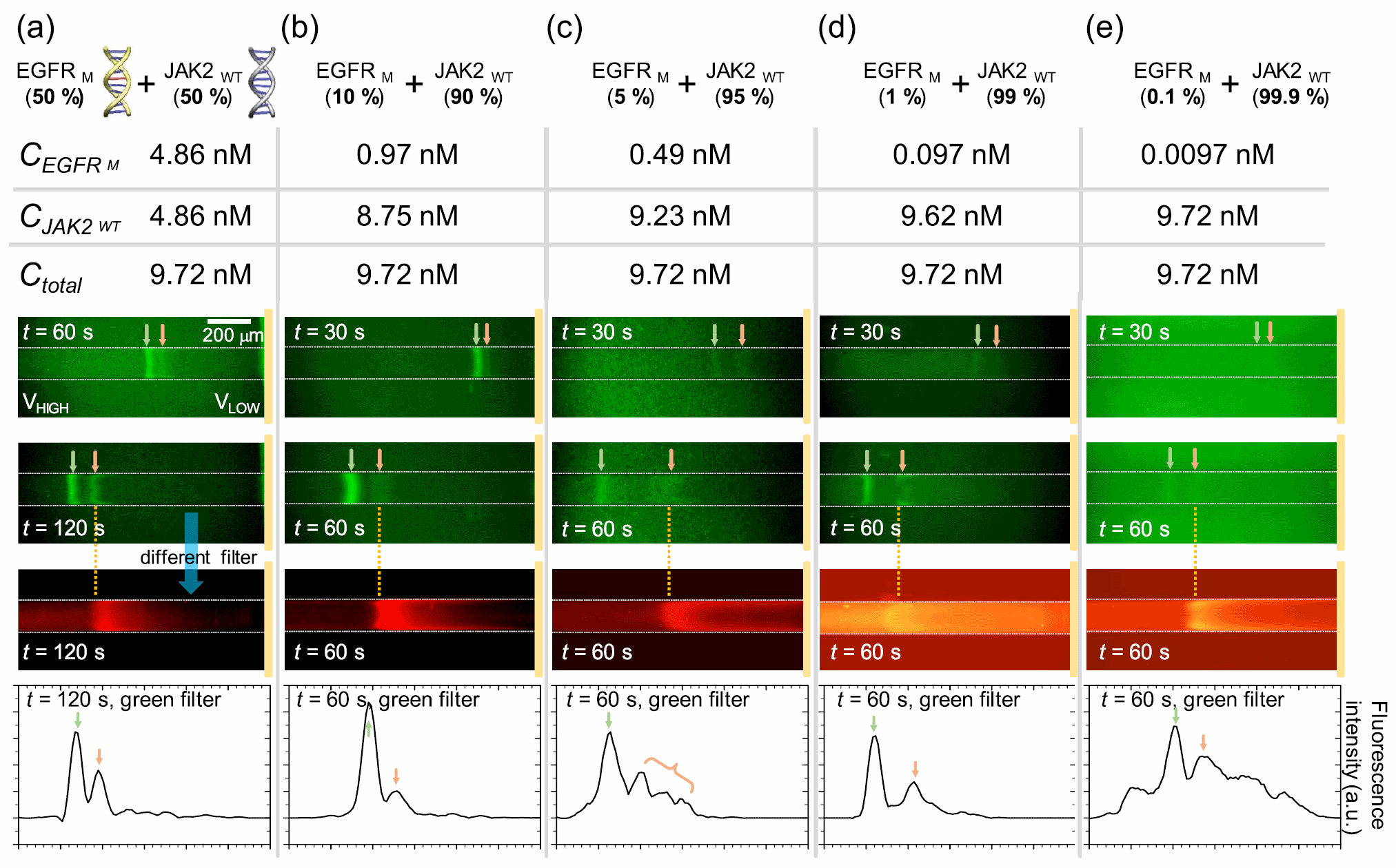
Figure 3. Experimental results on whether EGFRM can be detected in the stuff-DNA mixture. The ratio of target EGFRM and off-target JAK2WT was mixed as (a) 50:50, (b) 10:90, (c) 5:95, (d) 1:99, and (e) 0.1:99.9. In the case of panel (a), the applied voltage was 5 V for VLOW and 40 V for VHIGH. In all the cases except panel (a), the applied voltage was 5 V for VLOW and 70 V for VHIGH. For each experimental image, ISO, exposure time, etc. were adjusted for the presentation of the images. Thus, it can have similar fluorescence intensity even at different initial concentrations.
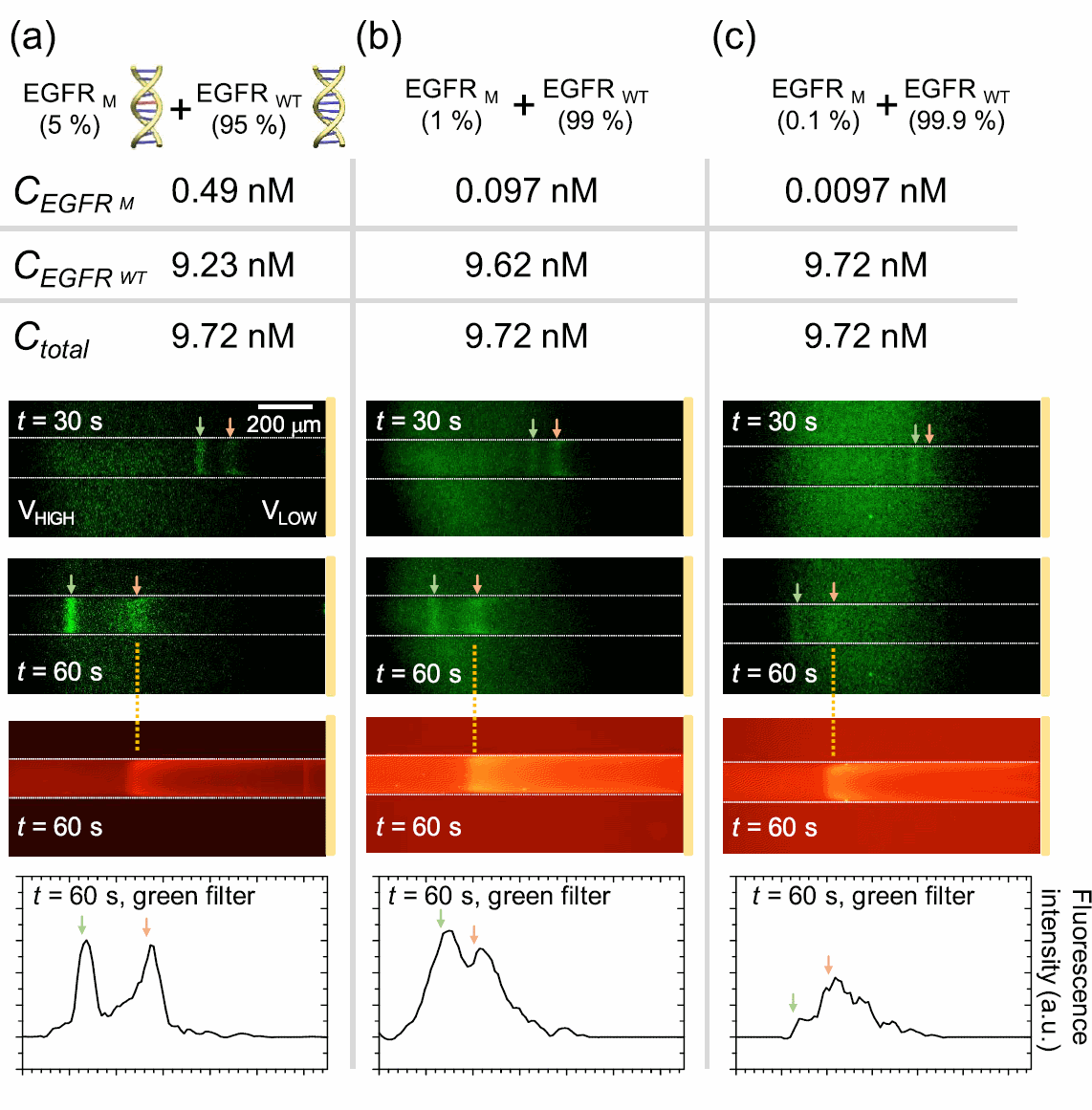
Figure 4. Experimental results on whether EGFRM can be detected in the heterologous mixture. The ratio of target EGFRM and off-target EGFRWT was mixed as (a) 5:95, (b) 1:99, and (c) 0.1:99.9. In all cases, two green plugs were observed in 1 min. For each experimental image, ISO, exposure time, etc. were adjusted for the presentation of the images. Thus, it can have similar fluorescence intensity even at different initial concentrations.